تم بحمد الله وفضله عمل ورشة عمل بوحدة الجهاز الهضمي جامعة الزقازيق وبالتعاون مع الزميل الفاضل اد.كريم عصام أستاذ الجهاز الهضمي بالقصر العيني الذي شرفنا بوجوده وخبرته الرائعةوالتي تم فيها عمل استئصال أورام من المعدة والقولون للمرضي وباستخدام منظار الموجات فوق الصوتية للتشخيص الدقيق مع أخذ العينات بالابرة القاطعة واستخدام تقنية الفراغ الثالث لاستئصال الأورام بنجاح، وكل الشكر والتقدير لرئيس القسم اد.عزت مصطفي وإدارة وحدة الجهاز الهضمي ولجميع اساتذتنا وزملاءنا وفريق التخدير علي دعمهم المستمروتعاونهم معنا والله الموفق.
فريق العمل:
اد.كريم عصام أستاذ الجهاز الهضمي بالقصر العيني.
اد.وسيم محمد أستاذ الجهاز الهضمي جامعة الزقازيق.
اد. هاني الصادق أستاذ الجهاز الهضمي جامعة الزقازيق
اد #سالم -يوسف أستاذ الجهاز الهضمي جامعة الزقازيق.
اد. عمرو شعبان أستاذ الجهاز الهضمي جامعة الزقازيق.
د.محمد عبد العظيم مدرس الجهاز الهضمي جامعة الزقازيق.
د.احمد محمود مدرس م الجهاز الهضمي جامعة الزقازيق.
Thanks to Allah’s grace and grace, a workshop was held at the gastroenterology and hepatology Unit, Zagazig University, in cooperation with our distinguished colleague, Dr. Karim Essam, Professor of Gastroenterology and hepatology at Kasr Al-Aini, who honored us with his presence and his incredible experience, in which the work was done.
Using endoscopic ultrasound(#EUS) for accurate diagnosis, taking biopsies with the needle before endoscopic submucosal dissection(#ESD).
Resection of tumors from the stomach and colon using the third space technique to excise the tumors successfully.
All thanks and appreciation to the president of our department, Prof. Ezzat Mostafa, the members of the gastroenterology unit, and all our professors, colleagues, and the anesthesia team for their support and continued cooperation with us.
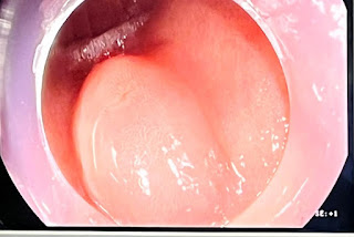 |
| fig1. gastric submucosal lesion |
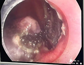 |
| fig2. ESD |
 |
| fig3. after resection |
 |
| fig4. gastric polyps |
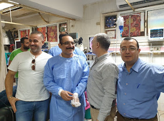 |
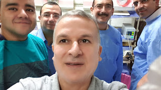
| fig5. Gastroenterologists and members of the team |
fig6. Gastroenterologists and members of the team
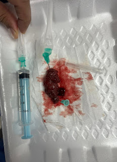 |
| fig7. lesion after resection |
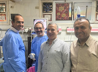 |
| fig8. gastroenterologists |
fig9. gastroenterologists
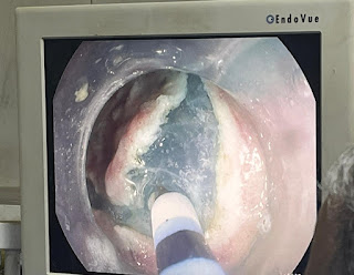 |
| fig10. knife resection |
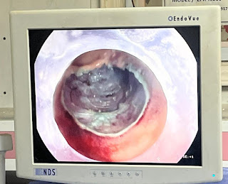 |
| fig11. bare area after resection |
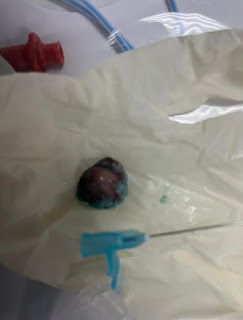 |
| fig12. mass after resection |
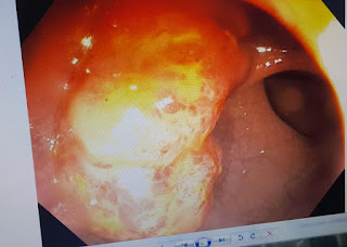 |
| fig13. Anal mass before dissection |

Comments