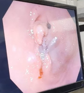
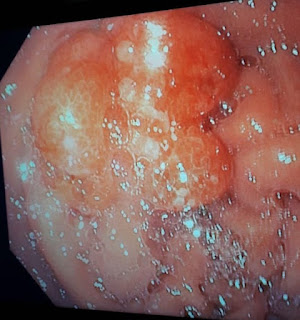
A 49-year-old woman presented with colicky abdominal pain was admitted to the endoscopy unit. The patient had a history of upper digestive endoscopy one year ago. The scope revealed a large antral polyp with overlying ulceration and bleeding. Multiple biopsies were taken, and histopathology revealed hyperplastic polyps. Computed tomography of the abdomen and chest is free. The patient underwent upper digestive endoscopy. We removed the polyp using the EMR technique with snare polypectomy.
Some tricks about this case:
1- Despite the polyp with ulceration and bleeding, there is no symptomatic melena or hematemesis. Also, the patient hemoglobin level is 14.6 gm/dl.
2- Despite it being a relatively sizeable lobulated polyp, the CT scan is free.
3- When carefully examining the stomach and the inflation of the stomach with air, the polyp disappeared. After reviewing the duodenum again, we found that the polyp prolapsed into the bulb. With minimal suction by scope, the polyp reappeared again in the antrum. It acts as a ball and valve syndrome.
4- When injecting methylene blue in the submucosa around the polyp, there is much resistance; it may be due to a- fibrosis from the previous biopsies b- fibrosis from prolapse of the polyp into the duodenum. C- it may be due to the long axis of the needle. We changed the injection needle and the endoscopist.
5- Cutting the polyp was done, but it was difficult to put the snare shaft at five o’clock, which may be due to the position of the polyp.
6- You can notice that the is excessive peristalsis during the procedure.
7- Histopathology revealed villous adenoma.
What is your opinion on the next endoscopy?
 |
| fig3 |


Comments