A 77-year-old man presented with melena and pallor (Hb= 7gm/dl)
was admitted to the endoscopy unit. The patient gives a history of child C
liver cirrhosis. The patient underwent upper digestive endoscopy. The scope
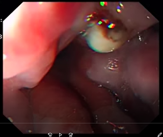
showed a fibrin plug with a surrounding reddish area on esophageal varices.
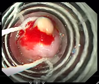
Just before the banding of this fibrin plug, this plug began to ooze. Banding of
the plug with 2 rubber bands was done. To make sure that the bleeding stopped,
the endoscopist checked the site of banding (video 3, also showed 2 variceal
bands that partially obstruct the esophageal lumen) and it looked good.
 |
| fig1: fibrin plug |
fig 2: banding of two esophageal varices, partially obstructing the esophagus
 |
| fig3: band ligation of a fibrin plug |

Comments